Using Orthopedic Braces for Healing Tendons, Joints and Bones in Dogs
Orthopedics is the branch of medicine and surgery that is concerned with the health of the bones and associated tissues. There are many types of orthopedic problems that affect dogs with and without additional disabilities. It would actually be unusual for a dog to live its entire life-span without experiencing lameness of one degree or another since they have 4 legs! Many of these incidents are minor problems that can resolve on their own with rest and time. Minor sprains, twists, or muscle bruises account for a large percentage of these common problems. Persistent lameness, any lameness associated with trauma, and severe lameness problems should always receive the attention of a veterinarian. Recovery from an orthopedic problem may take weeks to months and require excessive crate time without access to orthopedic splints and devices to aid healing.
Bandaging and applying splints is something of an artform. Injury of tendons, joints and bones in dogs often require surgery and long recuperation times. Often you are told by your veterinarian that the dog will require weeks or months of crate rest, which is often not easy with a young or active dog. Often splints and other orthopedic devices can lessen the crate time by giving the joint support while walking.
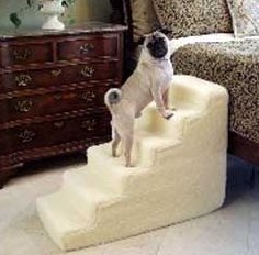
Unfortunately, joint-aggravating conditions like joint and mobility pain, hip pain, genetic defects, infections, and injury are all common, can all cause pain and impairment in dogs. All are tremendously hard for a loving owner to watch their dog struggle through. Difficulty with stairs, romping around the yard, jumping up to lay with you, or even simply walking at all can be extremely painful for our canine commpanions - their fearful avoidance of all these things are consequences of joint pain.
Which Splint Does Your Dog Need?
With all the dog braces out there, which one is right for your pet? Your dog may be a candidate for a dog brace or other mobility aid if he suffers from any of the following conditions:
- Arthritis
- Sprain or muscle weakness
- Hip dysplasia (mild to moderate)
- Back leg fatigue
- Post-operative or injury recovery
- Joint weakness or pain
- Limping
- Mobility handicap
Dog Braces Help Physical and Emotional Well-Being
Beyond drugs, utilizing a mobility aid can help restore your dog's confidence and enable pain-free activity. Many of the pharmaceuticals availalbe for dogs have side effects that compromise the kidneys and liver. If your dog can be helped with a physical device and avoid some of these poisons, wouldn't you do it? I would! Also, sometimes surgical intervention is needed to correct breaks, tears and general wear on dog joints - braces and splints can give supurb support after surgery or may even allow the joint to rest enough to prevent surgery. Dogs need to walk for their emotional well being, daily exercise and play are necessary. If he doesn't get what he needs, your dog can develop additional physical and emotional disorders, such as obesity, heart disease, bone disorders, anxiety and aggression.
A heartwarming story about a pony with leg braces.
Locating the problem area and potential cause
Limping or lameness is one of the more common medical problems encountered by dog owners. Often, the challenge comes when trying to determine the location and then the cause for the lameness. At first, it is important to consider the following questions. These are also questions that a local veterinarian may ask when evaluating the animal:
- What is the activity level of the dog?
- Any prior surgeries or medical problems?
- Does the problem get worse after exercise?
- Is the dog currently on any pain or anti-inflammatory medications?
- Has the condition come on suddenly or slowly?
- Does the problem seem to be associated with an injury?
- What is the animal’s age, sex, breed, size, and diet?
A thorough physical and orthopedic examination should then be performed with the goal of finding the cause(s) of lameness. The following are some basic steps that are often used when performing an orthopedic exam:
- The dog should be observed while standing. Signs of muscle shrinking (atrophy), confirmation abnormalities, swelling, and pain can often be found. The normal dog will bear 60% of its weight on its front limbs. When severe problems occur in the hind limbs, the dog will shift much of its weight to the front limbs.
- Then the dog is observed from the front, side, and rear while walking on a leash and at a slow trot. When the lame limb contacts the ground, the stride of the affected limb is usually shortened as compared to the opposite, normal limb. When the sound limb hits the ground, the animal will often spend more time with that limb in contact with the ground during the walking motion. Other problems that are often noticed include ataxia (lack of coordination), paralysis, paresis, and short, choppy gaits. If more than one limb seems to have a problem, realize that a perfectly normal gait requires the use of almost the entire nervous system and many of the muscles and bones of the body. Injury, damage, or tumors that affect the nervous system, muscles, or bones can cause problems in one, two, or all four limbs.
- Once the problem is identified, the veterinarian will do a thorough examination of the limb(s) is necessary to further localize the problem. He will feel feel every bone and every joint in the limb, including muscles, bones and tendons. All this should be done while the animal is fully awake without any sedation. Sedation may mask the animal’s response to the testing. Each joint is moved through the entire range of motion by flexing, extending, and rotating. Each joint should also be felt for evidence of swelling, pain, heat, lack of motion, instability, crepitus (popping), and laxity. Abnormal laxity (slackness) may indicate ligamentous injury or damage to the joint capsule. Some fractures are often very obvious, while others may only be identified by excessive movement of the area, pain, swelling, and lack of use of the limb. A hot, swollen joint often is the result of an infection causing inflammation. If the injury has caused the animal to not use the limb(s) for a period of time, the muscles may shrink (atrophy). Injury to muscles is often identified by tenderness, swelling, pain, and heat.
- After a specific area of injury has been identified, radiographs (x-rays) are often used to help determine the extent of the damage and determine the appropriate treatment. The cost involved in treating an orthopedic problem may be quite high, however, the outcome is usually good with good treatment and patience.
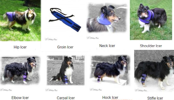
Hip Brace for Dog's Lower Back and Hip
A hip brace is designed to support the lower back and hip area. A brace of nylon, neoprene or other fabric wraps the lower back, hip and upper leg and is connected to a harness.
Hock Braces for Dogs
There are seven small bones in two rows that connect the lower hind limb to the paw and make up the tarsal joints. There is a separate joint connecting each row of bones. The calcaneus (heel bone) is the largest of these bones and projects back, toward the dog’s tail. The Achilles tendon attaches to this bone and travels up to the muscles on the back of the dog’s lower limb. The Achilles tendon is rather exposed and can be injured easily. Failure of the Achilles tendon also occurs due to fatigue. Injuries to the hock joints and bones are common in dogs. Most of these are soft tissue injuries, involving the ligaments and the joint capsule only. X-rays are always needed following injury to this area to help evaluate the small bones of the hock and to determine if any fractures exist. Fractures can also occur in any of the bones of the tarsus. Such fractures are not common, however, except in racing greyhounds that commonly suffer fractures of the heel bone. Dislocations of the tarsus can occur along any of the joints. If there is severe damage to ligaments and other structures that support the tarsus, fusion of the joint with a surgical procedure called arthrodesis may be necessary.
Dislocation of the tarsus can occur at any of the joints as a result of traumatic injury to the hind limb. Injuries to ligaments in the joint and sometimes fractures of the small bones are associated with hock dislocation. Diagnosis is made by careful examination by a veterinarian and x-rays. Replacement of damaged ligaments, repair of fractured bones, or fusion of the joint, also known as "arthrodesis," may be necessary to restore function.
Severing of the Achilles tendon can happen as a result of blunt trauma or sharp injuries to the hind limb. Fatigue failure can occur as well, resulting in swelling or even tearing of the tendon. Fatigue failure is caused by the gradual weakening of the tendon over months or years. This type of injury is more common in dogs that are vertical jumpers. Fatigue failure results in a tendon that has lost its "spring" and provides little support to the hock joint. Shortening of the tendon can be accomplished with surgery. If the tendon is torn, surgery is required to repair it. Some cases respond to splinting or casting the leg at its full stretched length, which helps the tendon to pull tight again as it heals.
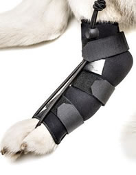
A dog hock brace used in conditions which cause lesions in the region of the hock joint, with symptoms such as pain, lameness or difficulty in moving. It is commonly used to support hock instability, collateral ligament, tendon rupture, osteoarthritis, tarsal arthrodesis, and joint fusion either as a support to surgery or instead of surgery.
Pet Stiffle or Knee Braces
The stifle joint is another hinge joint like the elbow but more complex. Stifle dislocation is a very serious problem where numerous ligaments and other tissues have been severely damaged. It requires a great deal of force to cause dislocation of the stifle joint. Dislocation of the stifle joint usually includes tearing and severe damage to both cruciate ligaments and at least one of the menisci. Diagnosis is made by careful examination by a veterinarian while the animal is under anesthesia and X-rays are very helpful as well, showing damage to parts of the joint that cannot be examined from the outside. Treatment is done by carefully reconstructing the joint through lengthy and very expensive surgery. If reconstructive surgery is successful and proper care is taken afterwards for healing, the outcome is often very good. Mild arthritis and/or stiffness of the joint may result eventually, however.
The lower portion of the femur ends at the stifle joint. On the front side of the lower end of the femur sits the patella (knee-cap), a small bone that moves up and down as the stifle bends. Small cushions of cartilage sit in between the lower end of the femur and the upper end of the tibia. These cushions of cartilage are called menisci (the lateral meniscus on the outside and the medial meniscus on the inside). Two ligaments run down the sides of the stifle joint, and two ligaments criss-cross inside the stifle joint. All these parts are important in the function of the stifle joint, and all can be injured and lead to pain and difficulty walking.
Injury to the stifle joint is common, and may involve fractures, ligament injuries, or dislocation. The most common injury to the stifle joint is the tearing of one of the inner criss-cross ligaments. This ligament is called the cranial cruciate ligament and is identical to the anterior cruciate ligament in the human knee joint. Improper movement of the patella or knee-cap.. Arthritis of the stifle is seen frequently in older dogs, usually as a result of some previous injury or problem in the joint. Growing bone diseases such as osteochondrosis as well as bone cancers may also affect the stifle area.
Cruciate Ligament Injuries
Cranial cruciate ligament injuries occur very frequently in dogs of all sizes and breeds, but large dogs seem to have it happen the most. When the cranial cruciate ligament (CCL) tears, this allows the tibia to slide forward away from the femur too much when the dog bears weight on the leg. This excessive movement stretches the other tissues in the stifle area,and is painful. The sliding movement also damages the cartilage cushions (menisci) that sit between the two bones. Most dogs with a torn CCL are very reluctant to put weight on the leg. The lameness is usually sudden, although many dogs randomly show signs for months before. Tearing can happen in an otherwise very healthy stifle joint with a sudden and severe blunt force to the area. Usually the ligament will gradually weaken and deteriorate as the dog ages. Aged, weak ligaments will often tear easily with minor force, such as slipping or jumping. Usually the owner will not know recall what happened to cause the injury. These ligaments age and become weak in both stifle joints over time, and when the CCL tears on one side, there is a good chance that the other will also tear eventually because extra weight stresses the "good" leg when one ligament tears. Overweight dogs are more likely to suffer CCL tears for the same reason.
Diagnosis of CCL tears is made during a detailed exam by a veterinarian. The forward movement of the tibia is detected and helps determine the diagnosis. X-rays can also be helpful in showing inflammation in the stifle joint but cannot show the actual torn ligament. Torn cranial cruciate ligaments should always be repaired with surgery. If not repaired surgically, the joint does stabilize itself the best it can with thickening of tissues. The dog will improve gradually up to a limited point after many months of lameness. However, arthritis will always result and is often severe. So if left alone and not repaired with surgery, the dog will usually appear to improve for awhile but will end up getting worse in the end. Once the arthritis sets in, it is irreversible. Prognosis with surgery is usually very good.
Patellar Luxation or Dislocation of the Knee-cap in Dogs
Patellar luxation (dislocation of the knee-cap) is another very common condition of the stifle joint of dogs. This condition occurs in both small and large breed dogs, but the type of luxation is usually a little different. The patella dislocates towards the inside of the leg in small breed dogs, while in large breed dogs, it usually dislocates toward the outside. The small breed version, known as medial patellar luxation (MPL), is more common.
Trauma to the stifle can cause dislocation a healthy dog. Patellar instability is graded based on its severity, Grade 1 is the most mild form, while Grade 4 is the most severe. Mild cases will gradually progress to become severe later on. Many dogs get used to the patella popping in and out of place as they walk.Some dogs this problem so well that the owner may not realize there is a problem until it is very severe. Arthritis of the stifle is usually a consequence of patellar dislocation.
Diagnosis of this problem is made primarily by physical exam. A veterinarian can detect the abnormal movement of the patella and determine the severity of the problem. X-rays are helpful with the diagnosis, especially in showing if any arthritis is present. Treatment depends on the situation. Mild cases with no lameness present are monitored for worsening of the condition. Severe cases require surgery. Treatment for cases falling in between must be judged individually by the owner and the veterinarian. Surgery can be extremely helpful for one dog but may not work at all in another dog. Small dog inside dislocation may respond very well to surgery, but success varies depending on the dog. Many large breed dogs have other deformities and so the success rate is not as good.
A dog knee brace is designed for cases of cruciate ligament, luxation of the patella, knee joint conditions such as arthritis and arthrosis, varus and valgus. It can be used both as an alternative to surgery, and in post-surgery.
Dog Carpal Braces
The carpal joint on a dog is comparable to your wrist as a human. The carpus is a very complex structure. There are seven small bones in two rows that connect the lower front limb to the paw and make up the carpal joints. There is a separate joint connecting each row of bones, making a total of three joints in the carpus. Injuries to the carpal joints and bones are common in dogs. Most of these are soft tissue injuries, involving the ligaments and joint capsule only. X-rays are always needed following injury to this area to examine the small bones of the carpus and to determine if any fractures exist. Fractures can occur in any of the bones of the carpus, although they are infrequent. Dislocations of the carpus can occur along any of the joints. If there is severe damage to ligaments and other structures that support the carpus, fusion of the joint (a surgical procedure called arthrodesis) may be necessary. At least one condition related to nutrition in puppies affects the carpus. The radius near its attachment to the carpus is a very common site for a very serious type of cancer known as osteosarcoma.
- Hyperflexion deformity is a condition seen in puppies 6-12 weeks of age. The Doberman pinscher breed is most commonly affected. The puppy stands abnormally on both front legs, with the paws seeming to knuckle under. Often, the condition improves on its own within a month or two. Exercise should be restricted, and the puppy should not be fed any nutritional supplements such as vitamin and mineral additives. Some studies show that this condition may be a result of "overnutrition."
Dislocation of the carpus can occur at any of the three joints as a result of traumatic injury to the front limb. Damage to the ligaments and other tissues that support the carpus is usually difficult to repair and does not heal very well. Special x-ray pictures can help to diagnose a dislocation at one or more of the three joints in the carpus. A dislocation of any or all of these is generally termed "hyperextension." When weight-bearing, a hyperextended carpus allows the paw to drop closer to the ground than normal. The condition is usually very painful. Minor cases of hyperextension of the carpus may be successfully treated with a splint or bandage and strict rest. Sometimes a hyperextension situation will require more aggressive treatment in medium to large breed dogs. Fusion of the joint, a surgical procedure known as "arthrodesis," is necessary in many cases to restore function.
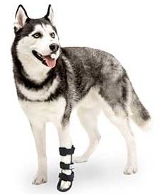
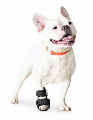
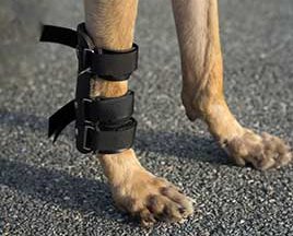
A carpal brace is designed to support the radiocarpal, carpometacarpal and tarsometatarsal joints. It is ideal for cases of hyperflexion or hyperextension (over extension) of the carpus and tarsus, ligament injury, sprains and fracture of the phalanges, metacarpal, metatarsal bones and when used diligently, may allow you to avoid surgery or even be preferable to surgery. A wrist brace or wrap is designed to stabilize the forearm bone, wrist and leg muscles and tendons. A brace of nylon, neoprene or other fabric is fastened an inch above the wrist bend.
Dog Elbow Braces
The elbow joint is a "hinge" joint connecting the humerus of the upper front limb to the radius and ulna of the lower front limb. The elbow is relatively unprotected and can be easily damaged and fractured. Dislocations of the elbow joint can also occur. Arthritis of the elbow joint occurs often in large breed dogs. A variety of growing bone diseases affect the elbow joint such as Osteochondrosis (OCD) - a growing bone disease affecting many areas of the skeleton. Bone and joint cancers can affect the elbow joint but seem to do so with less frequency than in other locations in the body.
- Fragmented medial coronoid process of the ulna (FMCP)is perhaps the most common cause of pain in the elbow joint of young large dogs. The ulna is a complex bone with several small projections at the hinge area where it connects to the humerus. Most of these projections develop separately in the young dog and attach themselves to their proper locations on the ulna as the dog matures. The medial coronoid process is one of these projections found on the inside surface of the elbow. A developmental "accident" occurs in which the process either attaches and then breaks off or fails to attach itself altogether. This small piece of bone "floats" around in the elbow, causing pain and inflammation. The fragment of bone is often too small to be seen on normal x-rays and may require arthroscopy (camera-assisted surgery) to diagnose. Treatment may be conservative, with anti-inflammatory pain killers and regular, gradually increasing exercise. Surgery to remove the fragment is also a possible treatment option. Arthritis is a common consequence of FMCP and may happen regardless of treatment.
Ununited anconeal process (UAP) is another growing bone condition affecting the elbow. The ulna is a complex bone with several small projections. The very top projection, which is part of the hinge mechanism itself, is called the anconeal process. The anconeal process normally grows separately from the main ulna bone in the very young dog, attaching itself at about 4-5 months of age. If the process fails to attach itself, it becomes a source of pain and inflammation in the joint. UAP is best diagnosed with x-rays Surgery is considered the best treatment option for most dogs, either by reattaching the process or removing it altogether. Arthritis often occurs later, but is helped by treatment. There is a genetic factor.
Elbow joint dislocation is not too common but can occur as a result of severe trauma. The joint is quite stable, and can result in a fractured bone more often than a dislocation of the elbow. X-rays are needed to tell the difference between fractures and dislocations but with severe injury, both can be present. Dog Elbow Braces are designed to protect elbows affected by hygromas, sores and calluses as well as fractures.. The brace is usually fitted with comfortable replaceable paddings and is helpful for conditions such as arthritis, arthrosis or elbow displasia.
Treatment of dislocations requires setting the bones in their proper positions while the dog is under anesthesia. If the procedure can be done within a few hours after the trauma, the setting of the bones can often be performed without surgery. The more time that has passed since the dislocation occurred, the more difficult it becomes to place the bones back in their normal positions. Surgery is necessary to set the bones if they cannot be replaced in their normal positions with anesthesia alone. Placement of a splint or bandage after setting the bones in place may be helpful to achieve rapid healing. Arthritis is common later in life in any joint that has been dislocated.
Front Leg & Shoulder Braces for Dogs with broken bones, Arthritis
The radius and ulna are paired bones that connect the elbow to the carpus or wrist joint. The radius is the major weight-bearing bone of the two. Fractures of the radius and/or ulna are fairly common in dogs. When fractures of the lower front limb occur, both bones are usually broken together. X-rays are usually not needed to know whether a fracture of the lower front limb is present but are important to determine the nature of the fracture. Many fractures require surgery, while others can be properly treated with a cast or splint to stabilize the fractured bone(s).Major orthopedic problems that commonly affect the front limb include fractures, ligament and tendon injuries, growing bone diseases, dislocations, infections, and cancers.The ulna radius brace is used for fractures of the front leg (Radius-Ulna and Radius-Carpus). It acts as a splint to keep the bones secure and provide extra support in lieu of a cast or after it is removed.
The shoulder joint is the connection between the scapula (shoulder blade) and the humerus (upper leg). Dislocations and fractures in this area do occur but are unusual in dogs because the shoulder is well-protected by surrounding muscles and other tissues. Arthritis can also occur in the shoulder joint. Inflammation of a tendon running through the joint and attaching to the biceps muscle is a rather common source of pain in the shoulder. This is called "bicepital tenosynovitis" and may occur because of excessive stress or other shoulder conditions. Growing bone diseases, especially osteochondrosis (OCD) are relatively common in the shoulder area. The shoulder is also a common location for a deadly type of cancer called osteosarcoma.
Bicepital tenosynovitis is a condition seen mainly in large breed dogs. It can occur simply as a result of excessive exercise or working stress on the shoulder, or it can be secondary to any other shoulder condition. Exact diagnosis of this problem is usually not necessary but may be obtained by x-ray using a dye injected into the joint. Treatment consists of rest and the use of anti-inflammatory pain killers. Surgery may be needed in the most severe cases.
Shoulder joint dislocation can occur as a result of trauma in dogs of any size or breed. X-rays are usually needed to tell the difference between a shoulder dislocation and a fracture. Treatment always requires professional help. Sometimes the dislocation may be successfully treated by moving the bones back into place and wearing a special bandage for 2 weeks. Often, however, surgery is a necessary part of the treatment.
Shoulder instability in the dog can be likened to “rotator cuff" injury in humans. The most commonly injured structure is the medial glenohumeral ligament. This injury presents as a mild to moderate lameness made worse after exercise. The injury can be caused by repeated stress in sporting and athletic dogs or by sprawling out with legs wide when slipping in senior dogs. Diagnosis is made by a veterinarian skilled in manipulation of the shoulder after ruling out more common causes of thoracic limb injury. A vest-like 'splint' provides support to your dog with medial shoulder instabilities. These types of injuries are commonly a result of an injury of the medial glenohumeral ligament and can be the result of a strain and/or tear. Shoulder injuries can present with your dog showing lameness and can range from moderate to severe. These injuries typically become significantly worse after extended exercise. Medial shoulder instabilities often occur in sporting and athletic dogs, and can sometimes occur in older dogs who slip when trying to get up from a slick floor. Medial shoulder injuries are often the result of repeated stress to the glenohumeral ligament.
Pelvic Injury and Fractures in Dogs
The pelvis transfers the weight of the hind end to both hind legs. The pelvis is divided into different areas. Weight is first transferred from the lower spine to bones on the right and left sides. These bones are called the ilia (singular = ilium). The ilia transfer weight to the hip joints. Connecting the right and left sides of the pelvis are two other bone sets known as the pubis and the ischium. The ilia, pubis, and ischium together make up the pelvis. The acetabulum is the socket-like portion of the ilium that connects to the femur.
The most common problem encountered involving the pelvis is trauma and injury. Almost 25% of all fractures involve the pelvis in dogs. Other problems that may involve the pelvis include dislocations, infections of the bone or surrounding tissues, and cancers.
Pelvic fractures are extremely common with trauma of nearly any type. Because of the rigid shape of the pelvis, fractures are always multiple and usually occur in sets of three. Pelvic fractures range from minor to extremely devastating. Because the right and left ilia are the main weight-supporting portions of the pelvis, fractures of these bones are usually very serious. Fractures of the socket-like acetabulum that houses the ball of the hip joint are also extremely serious and usually end up causing arthritis of the hip later in life, regardless of how well they heal. Fractures of the ischium vary in severity but are quite often allowed to heal on their own because they usually do not affect the animal’s ability to bear weight. Diagnosis of pelvic fractures is made with x-rays. Treatment depends completely on the situation. Many minor to moderate pelvic fractures can heal very well with strict cage rest and anti-inflammatory pain killers alone. More serious types of fractures, such as those that affect the hip joint or cause changes in the natural shape of the pelvis, should be treated with surgery. Surgery for pelvic fracture repair is extremely difficult and is always expensive. Anti-inflammatory pain killers and laxatives to help reduce pain of normal passage of bowel movements are important aspects of the treatment plan for many pelvic fracture victims.
Dog Hip Braces and Hip Dysplasia
Canine hip dysplasia is common in large and giant breed dogs. It has been diagnosed in small and medium breeds as well. Dogs with hip dysplasia are born with normal hip joints, but changes occur during development and aging. Gradual loosening of the joint with swelling, pain, and damage to joint tissues occurs. Months to years may pass, but the final result of hip dysplasia is excruciating pain and crippling in the hind limbs.Hip braces of various designs are for dogs suffering from hip dysplasia, hip dislocation, paralysis of the hind legs and Osteoarthritis. Usually there are metal inserts and a tensioning system that allows compression to maintain the hips in their sockets in lieu of surgery.
Dislocation of the hip joint is caused by injury to the hind end in dogs. Blunt force trauma causes the ball to pop out of the socket, tearing the ligaments and capsule that help keep the joint in place. The x-ray is an important piece of information for proper treatment because it will also show the veterinarian what type of dislocation has occurred. Treatment consists of getting the hip joint back together and keeping it there. This can be difficult to do. If action is taken quickly, the head of the femur may be put back into place by working the ball back into the socket. This is performed by a veterinarian while the dog is under anesthesia. Because more inflammation in the injured area actually "locks" the bones in their abnormal positions, this procedure becomes increasingly more difficult with the passage of time.
Neck Braces for Dogs
Injuries to the skull occur relatively infrequently and are usually associated with blunt trauma, such as an automobile accident. Fractures of any bone in the skull can occur, and depending on what organs or soft tissues are involved, these fractures can be very serious. Head trauma with severe brain or brainstem injury is usually fatal. Most other injuries to the mouth, nose, and face are treatable, although permanent disfigurement can result. Because the possibilities of injury types are endless, each type of head injury must be managed on an individual basis and under the care of a veterinarian.A neck brace for dogs is used when there are problems with the cervical vertebrae. A neck brace can support necks with Osteoarthritis, Discospondylitis, Vertebral Instability (Wobbler Syndrome), Intervertebral Disk Disease (IVDD) and other neurological issues and hernias.
Arthritis and Disabled Dogs
Arthritis simply means inflammation of a joint. Arthritis can be broken down into several different categories based on the type, location, and cause of joint inflammation. Arthritis can affect healthy as well as disabled dogs.
Joints can be freely mobile, partially mobile, or immobile. While inflammation can occur in any of these, the freely mobile joints and, to a lesser extent, the partially mobile joints are those most likely to create problems for the body when they do become inflamed. This is more likely to happen when previous injury or other degenerative factors are present, such as autoimmune disease.
The disk joints in between vertebrae in the spine are the most common partially mobile joints where problems develop in the dog. Intervertebral disk disease, where the cushion-like disks in between the bones of the spine become deformed and cause back pain and sometimes paralysis, is commonly seen in many breeds. Freely mobile joints include all the joints of the limbs such as the shoulders, elbows, hips, stifles (knees), and all the joints in the paws ("knuckles"). While the specific makeup of each of these joints differs.
A thin layer of cartilage overlying the bone in each joint provides for frictionless motion. A thick, tough wall called the joint capsule attaches to the bone on all sides and protects the delicate cartilage from damage. The joint capsule also holds in the sticky, clear lubricating fluid that is found inside the joint. This fluid is called synovial fluid and also helps with joint lubrication and frictionless movement.
The three broad categories of arthritis seen in dogs include degenerative arthritis (very common), infectious arthritis (uncommon), and immune-based arthritis (uncommon).
Degenerative joint disease or Arthritis in Joints
Degenerative joint disease is the medical term for the extremely common degenerative type of arthritis that occurs in a dog's joints. Specific types of DJD have already been discussed such as hip dysplasia. The main features of DJD are the wearing away of the cartilage and the formation of new bone at the edges. Degeneration of a joint can occur for many reasons: hip dysplasia, genetics, diet, and excessive exercise in a growing puppy can be contributing factors. Previous injury to a joint or nearby structures, infection, and age are also common reasons for this type of arthritis to develop. DJD often gradually gets worse with time and may become extremely severe and even disabling. Any freely mobile joint may become affected, but in dogs the hips, elbows, and stifles are probably the most likely to suffer from DJD. The most common reasons for these joints to develop arthritis because of previous injury or disease.
Signs of degenerative arthritis are pain: limping, stiffness, reluctance to exercise, protectiveness and even snapping when painful areas are touched. In severe cases, the dog may stumble or fall frequently, may slip easily on stairs or slippery surfaces, or may become unable to walk at all, if more than one limb is affected.
Because of the decrease in activity often associated with arthritis, dogs may become overweight. This creates a "vicious cycle," where the additional weight puts extra strain on the joints, which makes the arthritis worse. This causes the dog to exercise less or move even less, leading back to more weight gain, and so on.
Diagnosis is based upon physical examination and x-ray films. Treatment for degenerative joint disease is complex. Prevention is by far the most effective way of dealing with any type of arthritis. One very good example is the treatment for a torn cranial cruciate ligament in the stifle joint. Without surgery, the chance of moderate to severe arthritis developing in the joint approaches 100%. If surgery is performed soon after the ligament has torn, however, arthritis will still probably develop but will be mild. Once degenerative joint disease has developed it's too lated, prevention is not possible. One approach available for some types of arthritis is joint replacement surgery. Joint replacement surgery is an extremely difficult and expensive treatment, but it may offer the best possible result for some disabled dogs. Total hip replacement for hip dysplasia is available and may offer the best alternative treatment for dogs with limited mobility.. Other types of surgery may be helpful, such as joint fusion or joint reconstruction.
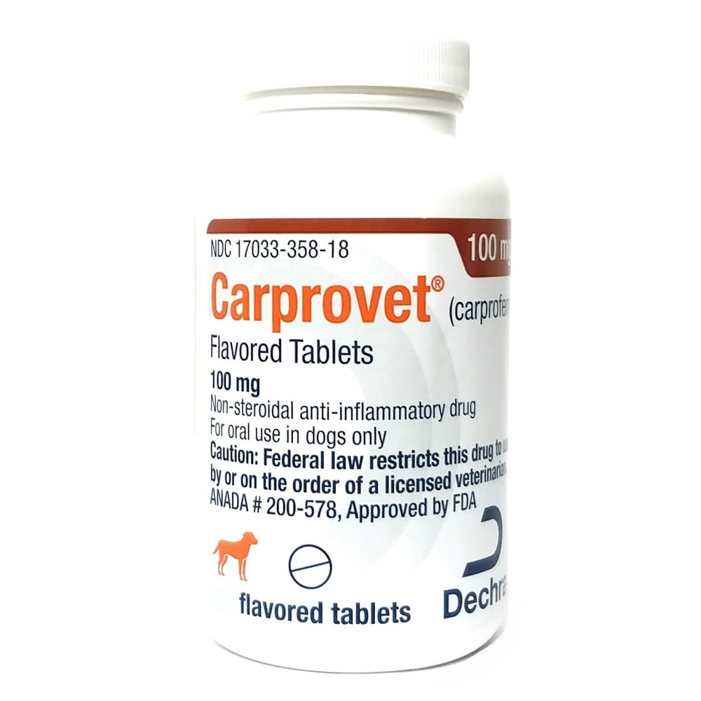
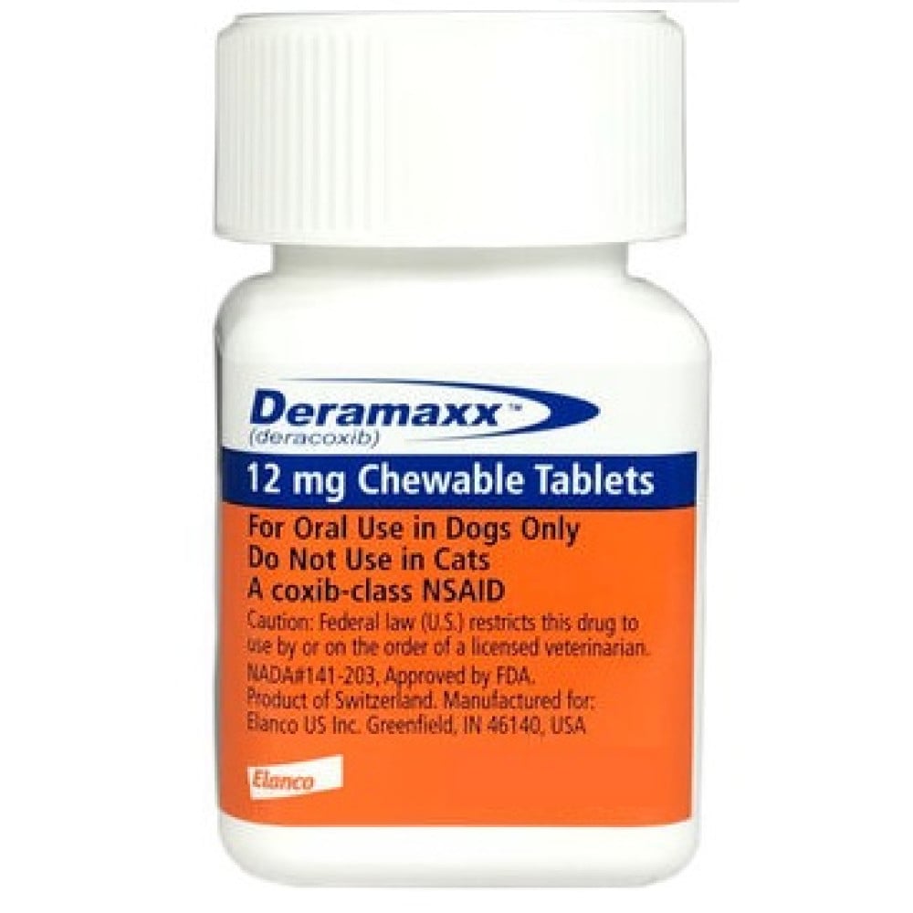
Most of the treatments available are for general pain relief. Another benefit of medical therapy is slowing down the progression of disease. There are two main groups of medications that are used to treat degenerative arthritis. The first group consists of anti-inflammatory drugs that help reduce pain and restore function. The second group consists of nutritional supplements containing natural hormones and other products thought to protect the joint tissues from damage and even change and improve the makeup of the joint tissues and fluid.
Get a High-Quality, Durable Brace
So as you can see, while surgery is an option most of the time - a brace can sometimes do either temporarily or even sometimes permanently. When shopping for dog braces and mobility aids, make sure to choose one made of durable material and quality construction. Check with the retailer about its exchange and return policy. You will want your veterinarian to show you how to put on the brace so it works optimally. Your first choice might not be the most comfortable for or acceptable to your dog. You will want to find a place that is willing to help you find what works best for your pet. If immobilization is the goal, so a break can mend or if there is a cut on a joint, you may want to invest in a splint. A splint allows muscles and tendons to rest, and tears to heal. It immobilizes joints so the joint can rest and heal.
Orthopedic problems are a common occurrence in dogs. The key to successfully treating these problems is early detection and proper treatment. The pet’s owner plays an essential part in identifying problems early and then seeking the proper medical attention. In general, if lameness in a pet lasts longer than 24 hours, or the lameness is getting worse - there is an obvious injury seek veterinary advice.
Common Conditions Where Dogs May Benefit from Splints
- Osteoarthritis of the carpal or metacarpal joints
- Degenerative Joint Disease (DJD)
- Polyarthritis
- Soft tissue injuries to carpal or metacarpal tendons/ligaments
- Carpal hyperextension
- Carpal or metacarpal joint instabilities or malformations
- Brachial plexus or radial nerve damage injuries
- Neurological conditions causing knuckling of paw
- Post-surgical protection
Hind Limb Conditions and Splints
- Osteoarthritis of the tarsal or metatarsal joints
- Degenerative Joint Disease (DJD)
- Polyarthritis
- Soft tissue injuries to tarsal or metatarsal tendons/ligaments
- Achilles tendon injury
- Tarsal hyperextension
- Tarsal or metatarsal joint instabilities or malformations
- Neurological conditions causing knuckling of paw
- Post-surgical protection
Caring for your Dog's Splint
- Do not allow your pet to chew on the splint. Use an Elizabethian collar if necessary.
- Splints must remain clean and dry to prevent moisture sitting against the skin.
- If the splint gets wet, allow it to fully dry before reapplying.
- It’s a good idea to check your pet’s toes daily for any swelling.If the toes become cool and/or puffy, it’s possible that the splint has been applied too tightly and is impairing the circulation of the limb and needs to be removed.
- If your pet develops chafing of the skin in any area due to rubbing of the splint, you may have to add some padding or baby powder to the affected area.
- As always, contact your veterinarian with any concerns.

